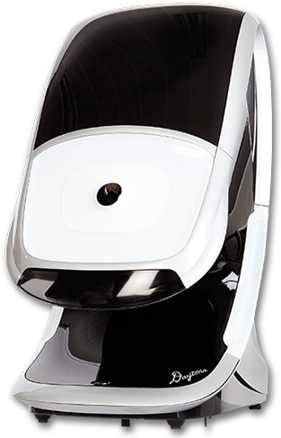
Optomap
We all want to to protect our eyesight and that is why it is important to have annual vision tests. This allows us to detect changes in the front of your eye so that alterations can be made to your eyeglass or contact lens prescription.. We also need to inspect the retina to check if it is healthy, damaged, or showing signs of disease.
When detected at early, many diseases can be treated without any further loss of vision. Each Optomap image is as individual as fingerprints or DNA and can provide us, and you, with a unique view of your health, quickly and comfortably. |
 |
| The Optomap image is captured in less than one second and is immediately available for review with you, to help you more understand the health of your eye. Because of the superior documentation ability of the Optomap™, we can monitor any condition more accurately as it progresses, and assist with treatment. It also gives us an accurate permanent record, from which we will have dedicated time to study, diagnose, and better treat your condition.
|
• More thorough/accurate exam
• Disease detection superiority
• Dilation not always necessary
• Monitor Incremental changes
• Ultra-wide field view of retina
• Very comfortable and quick
• Un-equaled documentation
• A non-invasive procedure
• Resume normal activities
|